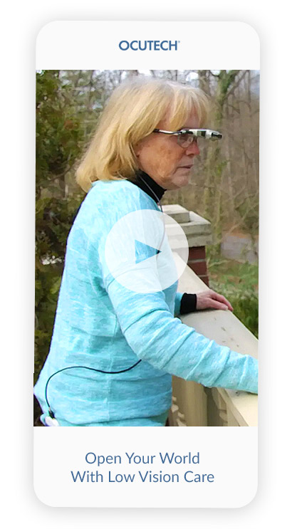
Ocutech bioptics can help you see beyond the limits of your Low Vision
Ocutech bioptic telescopes have helped many thousands of individuals throughout the world enhance their vision and enrich their lives.
Vision Disorders that Ocutech's Bioptics Can Help
Ocutech bioptics can help individuals with low vision from range of vision disorders including:
The retina, the light sensitive layer in the back of the eye, converts the eye's optical image into neural impulses transmitted to the brain along the optic nerve. The retina contains photoreceptor cells called rods and cones. Rods, which work most well in low light and are sensitive to motion, are primarily concentrated in the retina's periphery. Cones, mainly concentrated in the central macular area, are most sensitive to bright light and provide our color and detail vision.
Individuals who are born with achromatopsia (a lack of color vision), which is also known as stationary cone dystrophy and rod monochromatism, have cones that do not function properly, leaving them with reduced visual acuity, decreased color vision, and difficulty seeing in bright light. The degree of visual impairment can vary between “incomplete achromatopsia,” where some color vision is retained, and where visual acuity may be as good as 20/80, or “complete achromatopsia,” where none of the cones are functioning, there is no color vision, and visual acuity may be 20/200 or worse.
Individuals with achromatopsia often show mild to moderate changes in the macula. However, there may be no obvious changes in the retina's appearance early in the disease. Individuals with achromatopsia often have a pendular oscillating movement of the eyes called nystagmus. Reduced vision from achromatopsia does not worsen over time and do not lead to blindness. Researchers have discovered several genes that may cause achromatopsia; however, the CNGB3 is known to cause approximately 50% of cases.
How Bioptics Can Help Individuals With Achromatopsia
Individuals with this low vision disorder may benefit from using Ocutech bioptic telescopes to regain a significant amount of their ability to see clearly. They also find special red and magenta filters to be helpful both in and out of doors.
Albinism is a genetic defect that reduces the body's ability to produce or distribute melanin, a natural pigment that gives color to hair, skin, and the eye's iris. Individuals with albinism have reduced visual acuity due to a lack of pigment in the retina and the underdevelopment of the central part of the retina, called the macula.
Fortunately, individuals with albinism respond very well to low vision care. Bioptics can provide individuals with close-to-normal vision.
When the eye develops, the fetal neural eye structure wraps around and joins together, eventually becoming the bottom of the eye. If the two sides do not combine completely, there can be a gap in the iris (the colored part of the eye) called a coloboma. This congenital eye condition produces a keyhole-like appearance in the pupil. This incomplete closure can also affect the choroid, retina, and optic nerve. The disorder can occur in one or both eyes. It is estimated that coloboma occurs in 0.5 to 0.7 per 10,000 births.
Vision loss from colobomas may vary from mild to severe depending upon its size and location. Occasionally, other ocular malformations or disorders may be associated with coloboma, including microphthalmia (a very small eye), glaucoma, nystagmus, strabismus (turned eyes or crossed eyes), or blind spots in the visual field.
How Low Vision Treatment Options Can Help Individuals With Coloboma
While no medical or surgical treatment is currently available to treat coloboma, low vision aids, including bioptic telescopes, can provide significant functional gains. The magnification and illumination controls that Ocutech bioptics provide can readily enhance visual functioning for individuals with coloboma.
Diabetes interferes with the body’s ability to use and store sugar (glucose). The disease can cause damage throughout the body, including the eyes. In severe cases of diabetes Individuals may develop diabetic retinopathy which can severely affect vision. Individuals with diabetes need routine eye screenings to help safeguard their vision.
Diabetic retinopathy occurs when tiny blood vessels in the retina leak blood and other fluids. This causes the retinal tissue to swell, resulting in cloudy or blurred vision. The condition usually affects both eyes. The longer a person has diabetes, the more likely they will develop diabetic retinopathy. If left untreated, diabetic retinopathy can lead to blindness.
Treatment of diabetic retinopathy varies depending on the extent of the disease. People with diabetic retinopathy may need laser surgery to seal leaking blood vessels or to deter other blood vessels from leaking. Your eye doctor may need to inject medications into the eye to decrease inflammation or stop the formation of new blood vessels.
People with advanced cases of diabetic retinopathy may need a surgical procedure to remove and replace the gel-like fluid in the back of the eye, called the vitreous. Surgery may also be needed to repair a retinal detachment.
How Low Vision Aids Help With Vision Loss Caused by Diabetic Retinopathy
Vision loss caused by diabetic retinopathy and macular edema (swelling) can benefit from magnification, proper lighting, and increased contrast. Low vision specialists can help individuals with diabetic vision loss identify the appropriate equipment and techniques to help address specific visual needs and goals.
Glaucoma is an eye condition that damages the optic nerve, often caused by increased pressure within the eye. Over time, this can lead to vision loss, starting with peripheral vision and eventually affecting central vision. Since glaucoma-related vision loss is so far irreversible, testing to detect high pressures or changes in the optic nerve should be performed routinely.
Glaucoma-related vision loss can significantly impact daily life, making activities like reading, driving, or recognizing faces challenging. Low vision aids can offer valuable support for individuals affected by glaucoma.
How Low Vision Aids Can Help Individuals With Glaucoma
Innovative bioptic telescopes as well as other low vision devices can empower individuals with glaucoma to maintain their independence. These technologies are customized to individual needs, aiding in activities like reading, watching television, or recognizing faces. Your low vision specialist can assess your vision and seeing goals and recommend the most suitable devices to enhance your daily life.
In addition to the dry and wet forms of macular degeneration, many types of eye disorders can be called “macular degeneration,” including juvenile macular degeneration (Stargardt’s disease), macular holes, Best’s disease, and epiretinal membranes (macular pucker).
Low Vision specialists can prescribe magnification devices, special lighting, and unique methods to increase contrast to help people with central vision loss read, watch TV, recognize faces, and generally see more clearly.
A macular hole is a small break in the macula in the center of the eye’s retina. The macula provides sharp, central vision for reading, driving, and seeing fine detail. Most of the eye’s interior is filled with vitreous, a gel-like substance that fills about 80% of the eye and helps it maintain its round shape. The vitreous contains fine fibers attached to the retina's surface. The vitreous slowly shrinks as we age and can pull away from the retinal surface. In most cases, no adverse effects result from this natural aging process. However, some patients may experience a small increase in floaters, which are little “cobwebs” or light specks with a star-like appearance that seem to float about in your field of vision.
If the vitreous is firmly attached to the retina when it pulls away, it can tear the retina and create a macular hole, causing blurred and distorted central vision. It can also occur with other eye disorders, like high myopia (nearsightedness), injury to the eye, retinal detachment, and, rarely, macular pucker. The size of the hole and its location will determine how much a person’s vision is affected.
Is a Macular Hole the Same as Age-Related Macular Degeneration?
No. Although the symptoms are similar, macular holes and age-related macular degeneration are separate and distinct eye conditions. Both conditions are common in people 60 and over.
Is My Other Eye at Risk for a Macular Hole?
If a macular hole exists in one eye, there is a 10-15% chance that a macular hole will develop in your other eye over your lifetime.
How Bioptics Improve the Vision of Those With Macular Holes
Since the size of the macular hole is small and its edges are usually sharply defined, magnification can be very effective in helping individuals with macular holes see better. Optical and electronic magnifiers can be beneficial for reading and Ocutech bioptic telescopes can be very useful for helping individuals see better at a distance and when engaging in visual activities at arm’s-length and beyond.
People with severe nearsightedness (high myopia) are at greater risk for myopic degeneration. Myopic degeneration commonly occurs during young adulthood and can lead to a gradual decrease in clear vision. Occassionally in a small percentage of patients vision can decrease more abruptly.
Although central vision may be lost, side (peripheral) vision usually remains unaffected. The causes of myopic degeneration are not clearly understood, but they may include anatomic or hereditary factors.
Fortunately, your remaining sight can still be useful, and with the help of low vision devices, people with myopic degeneration can continue to pursue and enjoy many of their everyday activities.
How Bioptics Allow Individuals With Myopic Degeneration To Lead Normal Lives
Vision loss caused by myopic degeneration responds very well to magnification. Ocutech bioptics can be especially helpful for seeing at a distance and make reading, seeing signs, faces, the computer, TV, and even driving a possibility.
Nystagmus is a term that describes the fast, involuntary movements of the eyes (fluttering) that can be side-to-side (horizontal nystagmus), up and down (vertical nystagmus), or rotary (rotary or torsional nystagmus). The term “dancing eyes” has been used to describe nystagmus. Perhaps the most famous individual with nystagmus is flutist James Galway.
There are two forms of nystagmus:
- Congenital Nystagmus: Individuals are born with this condition.
- Acquired Nystagmus: Develop later in life due to disease or injury.
Nystagmus eye movements can either be pendular (roughly equivalent speed in each direction) or jerk (where there is a fast movement in one direction and a slower recovery movement in the opposite direction). Pendular nystagmus is usually found in congenital nystagmus, while jerk nystagmus is present in acquired nystagmus.
Improving a Nystagmus Patient’s Quality of Life With Distance Vision Aids
Individuals with nystagmus most always have some reduction in visual acuity- ranging from a mild to a marked reduction. They usually also have high amounts of astigmatism and can also be farsighted or nearsighted. Even when glasses are prescribed, visual acuity does not return to normal.
Telescopes (monoculars) and versions mounted into eyeglasses (bioptic telescopes) can make distance vision very close to normal, allowing individuals with nystagmus to read signs while shopping, travel, play music and board games at normal distances and maybe even drive. Children in school can benefit greatly from bioptics allowing them to see their teacher and classmates, Nystagmus does not usually interfere with the effective use of telescopic devices.
Optic nerves, which contain 1.2 million fibers each, carry the neural impulses from the retina to the brain to enable us to see. Disorders of the optic nerve are caused either by developmental (genetic or abnormal development) or acquired factors (trauma or disease).
Optic nerve disorders can impact vision in a number of different ways and can affect one or both eyes. It can reduce how clearly the individual sees, it can reduce color vision and contrast, and it can affect different parts of the visual field.
How Low Vision Care Can Help Patients With Optic Nerve Disorders
Low vision care can be very effective in helping individuals with optic nerve diseases lead more normal lives. Vision loss from optic nerve disease usually responds well to magnification, and bioptics can help make watching TV, recognizing friends and family, viewing details on a computer, seeing street signs and even driving possible.
Retinitis pigmentosa is a disorder of the rods. It usually develops slowly and reduces night vision and the ability to see to the side (peripheral vision), sometimes called 'tunnel vision.' Individuals with retinitis pigmentosa can often read and walk during the day. Devices that expand the visual field, like image minifiers and field viewers, can help individuals with tunnel vision see better to the side, helping with mobility and other activities. Other disorders including Choideremia and late stage Glaucoma can cause similar vision complications.
A normal visual field of each eye extends to approximately 80 degrees nasally (to the nose) and 90 degrees temporarily (to the ear). A normally sighted individual can see an almost 180-degree field of view with both eyes together. (To understand what a degree is, extend your arm fully at shoulder height and bend your hand upward to the ceiling at the wrist. The width of your hand is approximately 10 degrees in diameter.)
Individuals with tunnel vision from disorders like retinitis pigmentosa will see more poorly at night and tend to bump into objects (for example, door jams and low-hanging branches). Since tunnel vision usually develops slowly individuals can learn to scan across txheir visual field to help them walk and move safely. When fields of view become very narrow (perhaps 10 degrees diameter or less), sighted guides, long canes, and guide dogs become very helpful.
How Ocutech's Tunnel Vision Aids Can Help
Ocutech makes three versions of optical minification devices: the Image Minifier, the Field Viewer, and the Field Expander. Compared to reversed telescopes that produce barrel distortion and an unnatural image, Ocutech minification systems are specially designed to provide a sharp, flat image that is more natural and easier to use.
Retinopathy of prematurity (ROP) is an eye complication affecting premature babies who have received intensive neonatal care in which oxygen therapy is used to support the premature development of their lungs. It is thought that the high amounts of oxygen required causes unnatural growth of the retinal blood vessels, which may result in scarring and retinal detachment. Vision complications from ROP canrange from mild to marked.
Low Vision Care Can Help Those with Retinopathy of Prematurity
Like most eye conditions that affect visual acuity, optical magnification aids, including Ocutech bioptics offer magnification options to address the full range of visual needs. Since ROP is present in childhood, early low vision aid support can be very helpful to maximize the child's learning, social and emotional development.
Initially described by German ophthalmologist Karl Stargardt in 1901, Stargardt’s disease, also known as fundus flavimaculatus, is a retinal disorder that affects the macula early in life and is often also called juvenile macular degeneration or early onset macular degeneration.
Stargardt’s disease does not affect the peripheral retina, and, as a result, individuals do not lose all of their vision. They can usually walk and engage in general activities with little difficulty. However, reading and seeing details, such as signs and faces at a distance, are reduced.
Ocutech bioptics are very popular among individuals with Stargardt’s Disease as they allow them to see better and live more normally.
How Ocutech's Tunnel Vision Aids Can Help
Ocutech makes three versions of optical minification devices: the Image Minifier, the Field Viewer, and the Field Expander. Compared to reversed telescopes that produce barrel distortion and an unnatural image, Ocutech minification systems are specially designed to provide a sharp, flat image that is more natural and easier to use.
How Ocutech Bioptics have Changed Lives!
“I could now see and do things I couldn’t ever do before. The Ocutech bioptics have literally changed my life.”
- Cody Smith
“I have regained my independence and drive, which has helped me train harder and harder to now be one of the best visually impaired competitive surfers in the world.’
- Aaron Paulk
“As soon as Lane put on the Ocutech bioptics, his face lit up! We’re just thankful to Ocutech for creating these aids that have been so helpful for our son.”
- Julie Hoskins
Can an Ocutech Bioptic Help Me?
If you can answer YES to the following four questions, then Ocutech bioptics may be helpful to you:
- Is your vision loss due to macular degeneration or a similar disorder?
- Is vision in the better eye with your prescription (if used) 20/300 (6/90) (0.07) or better?
- Can you watch TV or recognize people’s faces two feet away?
- Can you read headlines in a newspaper?
Our Self-Assessment Questionnaire can help you determine which Ocutech bioptic might be best for you.. Our qualified team will review your responses and provide a personalized report that you can take to your low vision specialist to help you explore the benefit of Ocutech bioptics.
It's up to you to take the next step to enhance your vision
Ready to embark on your journey to clearer vision? Ask your low vision specialist for an Ocutech bioptic evaluation, or contact us for a referral to your nearest provider.
By registering your Ocutech bioptic you’ll be able to receive updates on enhancements, new products, and new ideas from Ocutech.
Your story can matter! By sharing your experiences, you can inspire others considering an Ocutech bioptic to learn how your bioptic has impacted your life. If we can use your story, we'll offer you free routine maintenance services on your bioptic (a $100 Value).
FAQ Hub: Your Vision Questions Answered
Ocutech does not determine the cost of its products to consumers.
Ocutech products are ordered by prescribers who in turn prescribe and dispense them to their patients. The cost of Ocutech products may be organized into three categories:
- The base cost of the device.
- The cost of customization charges for eyeglass prescriptions, eyepiece correction, special filters, etc.
- The professional services associated with the evaluation, prescribing, fitting, and training of the individual using the device.
Benefits for low vision aids vary between insurers. You should contact your HR department or insurance company representative to confirm your coverage. VA Medical Centers do provide Ocutech products for their clients. Medicare does not presently cover low vision aids or appliances of any kind. Employers through the ADA Act may provide Ocutech products for their employees.
When billing insurance for the purchase of low vision telescopic aids, use the V2615 ophthalmic product code.
Ocutech’s frames have been specially designed or selected to meet the demands of incorporating Ocutech’s low vision aids. Most VES and SightScope bioptic telescopes must be used with frames provided by Ocutech. The VES®-Mini and the InstaMount can be attached to other suitable frames.
Ocutech’s VES systems are available in 3x, 4x, 5x and 6x powers. The SightScope Series is available in 0.5x (for tunnel vision), 1.7x, and 2.2x powers. Not all systems are available in all powers.
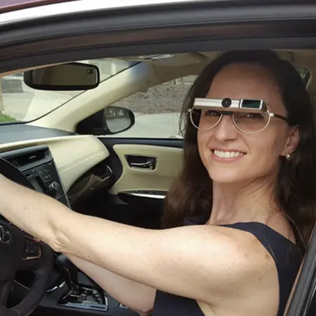
Ocutech bioptics can help visually impaired individuals to see television, movies, theater, faces, signs, blackboards, and classmates in school, shopping, travel, table-top activities including card and game playing, and mid-range activities such as reading music and the computer. Ocutech users have even used their systems to drive*, hike, golf, bowl, paint, fish, and mow the lawn.
*Bioptic telescopes may be used for driving in states where legal and with appropriate licensure.
Low vision specialists are trained to determine important technical details needed to properly order Ocutech low vision aids. We are happy to assist your prescriber in placing your order or to refer you to an Ocutech prescriber closest to you.
Ocutech warrants each of their products against defects in manufacture. The specific warranties vary and are contained on the individual technical sheets for each product.
Yes. Most homeowner’s insurance policies will allow you to add your Ocutech system as a rider much like they do for cameras and jewelry. Contact your insurance broker for additional information.
When billing insurance for the purchase of low vision telescopic aids, use the V2615 ophthalmic product code.